In and ex vivo imaging
Main Fields of Research
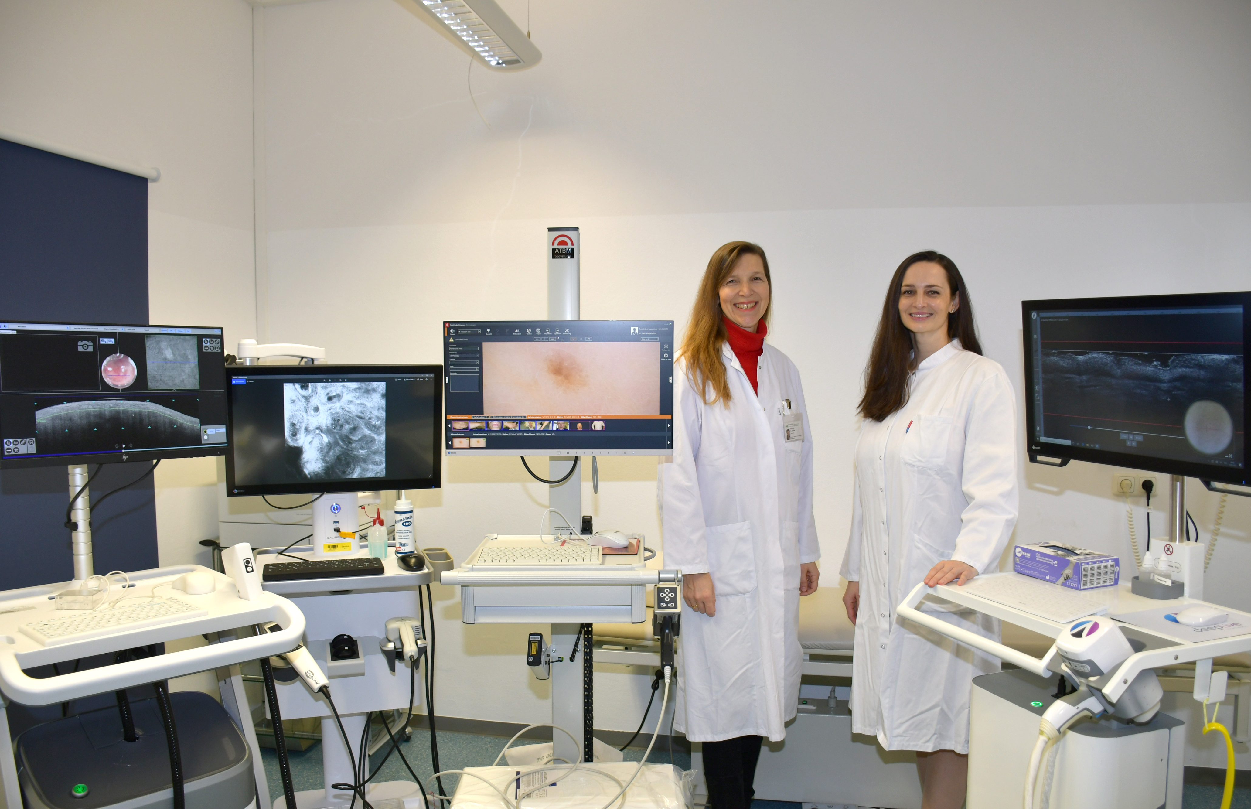
The main focus of the in vivo imaging group is to establish, develop and use modern non-invasive imaging techniques like optical coherence tomography, confocal laser microscopy and line-field confocal OCT to optimize the detection and early diagnostics of melanoma and non-melanoma skin cancer, but also for inflammatory and infectious dermatoses. In new prototype devices, we recognize and establish new patterns for diagnosis to improve sensitivity and specificity of the clinical diagnosis and correlate these findings to ex vivo imaging and histopathology results.
These methods are non-invasive without pain, surgery, local anaesthesia, scarring etc. but yet allow a precise diagnosis in real-time and therefore can spare unnecessary biopsies or excisions or speed up referral to the right treatment. As the same skin site can be visualized over time e.g. before and after treatment, these methods are also valuable tools for therapy monitoring.
Ex vivo confocal laser scanning microscopy (ex vivo CLSM) is a novel diagnostic method allowing rapid, high-resolution imaging of freshly excised skin samples in an intraoperative setting. It enables the surgeon to know the diagnosis and whether the resection edges are clear or not within few minutes after the excision. This innovative bedside histology may decrease the number of surgical procedures and length of hospitalization in the near future.